
In some unborn children, routine ultrasound examinations reveal a closure of the bladder (LUTO) and/or the upper urinary tract, i.e., the ureters and/or the renal pelvis and calyx system (UUTO).
This malformation prevents urine from flowing freely into the amniotic cavity. As a result, the bladder enlarges (megacystis). In severe cases, the bladder alone occupies a large part or even all of the child’s lower abdomen. In longer-standing cases, enlarged renal pelvises and ureters as well as conspicuously bright kidneys are also seen on ultrasound.
In cases of severe obstruction, both of the child’s kidneys can become so damaged within a few weeks that they stop producing urine long before birth. If this happens before the 22nd week of pregnancy, the lack of amniotic fluid (fetal urine) also causes life-threatening lung development problems in the unborn child (lung hypoplasia). Therefore, after a prenatal diagnosis, you should ideally go to a specialist center the following day.
In cases of severe congestion, the bladder may also rupture (bladder rupture). This presents as a massive accumulation of fluid in the fetal abdominal cavity (urine ascites), typically accompanied by a slightly enlarged bladder.
Similarly, in cases of severe prenatal urinary retention, one or both kidneys may rupture (renal rupture). This presents as a massive accumulation of fluid in the renal capsule on the affected side. Fetuses diagnosed with urinary ascites or a ruptured kidney should be referred to a specialist center immediately. Even these extreme cases can often be treated successfully there.
In less severe cases, the kidneys only lose their function in the last trimester of pregnancy. Since sufficient amniotic fluid is usually produced for lung development in these cases, life-threatening underdevelopment is less likely.
Fortunately, there are also milder cases with smaller bladders (<15 mm in diameter) that occur in early pregnancy and then regress spontaneously. However, it is advisable to rule out the presence of additional malformations of other organ systems and, in particular, genetic disorders!
Consequences of moderate and severe fetal urinary outflow disorders
Without prenatal therapy, most children with severe bladder outflow disorders and many children with moderate disorders lose their kidney function. In the group of children affected by severe outflow disorders before the twentieth week of pregnancy, lung function is also lost during the course of pregnancy without prenatal treatment.
Diagnosis
With the help of fetal ultrasound (sonography), it is easy to diagnose a therapeutically significant urinary retention if visibility conditions are adequate. We recommend treating this immediately. If possible, amniotic fluid or placental tissue can be obtained at the same time for genetic testing.
Fetal urine tests are not suitable for predicting later kidney function, especially in early pregnancy.
Due to the small size of fetal structures, the displacement of other internal organs by the enlarged urinary tract, and uncertainty in determining the sex, it is not possible to rule out other malformations with certainty, especially in early pregnancy.
Unfortunately, after very early diagnosis of severe obstructions around the
12th week of pregnancy, many examiners miss the therapeutic window of approximately 4 to 6 weeks for kidney function, for example by hoping for a possible spontaneous regression of the urinary retention and waiting for genetic testing to be carried out and the results to be available – or even waiting for a decrease in the amount of amniotic fluid, repeated punctures to determine urine values, etc.
At the same time, inexperienced examiners are usually unaware that many of the selection criteria proposed in the medical literature for prenatal interventions are not applicable to particularly affected children, especially in early pregnancy.
This in turn wastes valuable time for saving the kidneys until a correct diagnosis can be made.
It is crucial that you contact a specialist center immediately after your child is diagnosed with a urinary tract obstruction!
Treatment at the DZFT
Our center is one of the few in Germany that specializes in prenatal interventions for unborn babies with severe urinary outflow disorders.
Within the first few days after diagnosis, we at the DZFT begin to relieve the urinary retention in your unborn child.
Ideally, this should be done in the early stages of pregnancy. However, kidney function can still be preserved later in pregnancy if the retention develops late and has been present for less than 4-6 weeks! If possible, the material required for genetic diagnosis is obtained at the same time as the intervention.
After relieving the urinary tract, it is usually possible to determine the sex of the baby using ultrasound. This means that the trauma to the mother and child from the intervention and diagnosis is identical. All of the mother’s options regarding the further course of the pregnancy remain unaffected after these initial measures. In our opinion, the following are important advantages of our strategy for severe urinary outflow obstruction.
In selected cases and if the position of the fetus is favorable and the fetus is already large, the outflow obstruction can also be removed endoscopically by opening small, valve-like flaps in the fetal urethra using laser energy.
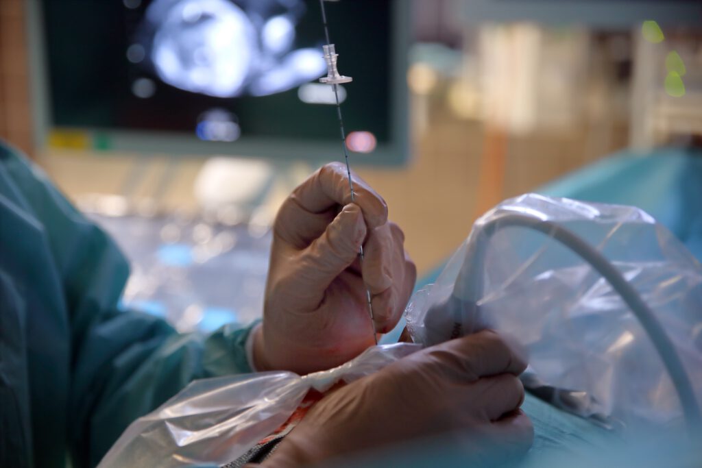
In most cases, only a small silicone tube or catheter is inserted into the bladder. The latter drains urine from the bladder into the amniotic cavity. This method is known as a vesico-amniotic shunt (VAS).
If the bladder diameter is less than 2.5-3 cm (figure – early LUTO), we usually use a small wire mesh (Somatex Intrauterine Shunt) in the early weeks of pregnancy when amniotic fluid is still present. In about one third of cases, when this shunt is used, a new shunt must be inserted during the course of pregnancy, as it is only about 3 cm long and can become dislodged as the fetus grows. We therefore recommend follow-up checks every 7–10 days until birth after insertion of this shunt so that any renewed accumulation can be relieved immediately.
If the bladder diameter is greater than 3 cm and the amount of amniotic fluid is already reduced, we prefer to place a long silicone catheter in the bladder. With the technique used at our center, it is not necessary to fill the amniotic cavity (amnion infusion). Due to the catheter being up to three times longer than the stent shunt, which is just under 3 cm long, a shunt system is usually sufficient for the duration of the pregnancy until birth.
This technique, developed at the DZFT, minimizes the manipulation required for catheter insertion. Since the outer limb of the catheter used cannot become caught in the uterine wall or placenta, as is the case with the conventional treatment using a double pigtail catheter or a wire stent shunt, the risk of premature rupture of the membranes is reduced. Since the drain also has a larger diameter and is significantly longer, it is less likely to become blocked or dislodged.
To perform the respective minimally invasive procedures, only a single incision of approximately 5 mm in length is usually required on the abdominal wall. For drainage, a needle with an outer diameter of only 1.2 mm is then inserted through the uterine wall into the amniotic cavity and then through the fetal abdominal wall into the bladder. When the silicone catheter is placed, the opening is dilated to just under 2.5 mm.
In some unborn babies, despite successful drainage of the bladder, significant congestion of the upper urinary tract (UUTO) and the kidneys (hydronephrosis) develops. Depending on the severity of the findings, we also treat these unborn babies in a second procedure, as severe obstruction of the upper urinary tract alone can, in principle, have the same negative consequences as LUTO (loss of kidney function, oligohydramnios, lung development disorder).
Results of shunt treatment at the DZFT – Time is kidney!
Approximately 4 out of 5 patients treated before the end of the 16th week of pregnancy and who survive the pregnancy are born with normal kidney function and functioning lungs after prenatal therapy.
In children treated from the 17th to the end of the 24th week, normal kidney function according to laboratory tests is still observed in 4 out of 10 children after birth.
If a urinary tract obstruction is only discovered in the 20th week of pregnancy, it is important to estimate how long the obstruction has been present – for more than 6 weeks or only recently. Pregnant women can often provide information on this. In all other cases, we endeavor to make our own accurate assessments by means of an ultrasound examination at our center. It is important to determine the timing for the prognosis of the child. We therefore strongly recommend referral to a specialist center.
Our latest studies show: In children born with normal kidney function according to laboratory tests, this function remains stable, at least during the first years of life, without significant deterioration!
Most children with LUTO can be saved through prenatal therapy, but ……
about a quarter of children diagnosed and/or treated prenatally with severe narrowing of the urinary tract do not survive pregnancy. Some unborn babies die before intervention. This could be due to excessive pressure and traction from the rapidly growing bladder on blood vessels in the fetus and along the surface of the bladder (umbilical artery, umbilical vein, aorta, vena cava). However, the most common reason for non-survival is the mother’s decision to terminate the pregnancy if amniotic fluid does not form after a technically successful procedure or if it is lost during the course of the pregnancy. A few unborn babies also die within 24 hours of intervention. Here, too, we suspect that problems with the care of the child are the cause.
In male fetuses, the most common cause is small valves in the urethra (posterior urethral valves), which act like a closed valve and block urine flow. Sometimes, however, the urethra is not formed (urethral atresia). The consequences for the kidneys are the same, but there are differences in postnatal treatment, which we will be happy to explain to you.
Female fetuses with urinary outflow disorders often have associated malformations throughout the entire urogenital area. Postnatal treatment in Germany is only available at a few specialized centers. At our center, we work closely with renowned specialist Prof. Dr. Raimund Stein: Info
If you would like to read about our treatment strategy in a scientific publication, please use the following link: Therapy at the DZFT
Further information about this and other life-saving treatments can be found here.
Patient comfort and safety are our top priorities
All prenatal shunt procedures in unborn babies with severe urinary tract obstructions are performed at the DZFT between the 12th and 32nd week of pregnancy in an operating room under short anesthesia with antibiotic prophylaxis.
Thanks to these measures, the technical success rate at our center is well over 90%, even in very difficult cases. No procedure-related infections have been observed. In addition, bladder rupture, complications in the child, and infections are less common. The stress caused by the procedure for both mother and child is also reduced to a minimum. In our opinion, this is an indispensable advantage, especially in the case of repeated procedures.
As incredible as it may sound, pain management for mothers and children and infection prevention through procedures performed in a dedicated operating room with the administration of antibiotics are still not standard practice for prenatal procedures in Germany! Please consider these important factors when choosing your treatment center!
If you have any further questions about minimally invasive treatment of prenatal urinary tract or kidney diseases or would like to obtain a second opinion, please contact the DZFT daily between 10 a.m. and 5 p.m. on 0175/597-1213 or send us an email.
If you only reach the answering machine, please leave your name and a telephone number where we can call you back.
If you have already decided to have a procedure performed at another clinic, we will be happy to advise you in this case as well and offer a prognostic second opinion on your child’s condition and treatment.
Please note: The content of our website is for informational purposes only and is not intended to replace personal consultations with us or other recognized medical specialists in this field. For all individual questions and decisions concerning your health and that of your child, we strongly recommend that you and your family members contact us, your doctor, and/or other experts in person.